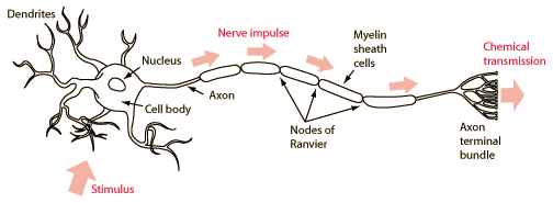
Nerve CellMany nerve cells are of the basic type illustrated above. Some kind of stimulus triggers an electric discharge of the cell which is analogous to the discharge of a capacitor. This produces an electrical pulse on the order of 50-70 millivolts called an action potential. The electrical impulse propagates down the fiber-like extension of the nerve cell (the axon). The speed of transmission depends upon the size of the fiber, but is on the order of tens of meters per second - not the speed of light transmission that occurs with electrical signals on wires. Once the signal reaches the axon terminal bundle, it may be transmitted to a neighboring nerve cell with the action of a chemical neurotransmitter. The dendrites serve as the stimulus receptors for the neuron, but they respond to a number of different types of stimuli. The neurons in the optic nerve respond to electrical stimuli sent by the cells of the retina. Other types of receptors respond to chemical neurotransmitters. The cell body contains the necessary structures for keeping the neuron functional. That includes the nucleus, mitochondria, and other organelles. Extending from the opposite side of the cell body is the long tubular extension called the axon. Surrounding the axon is the myelin sheath, which plays an important role in the rate of electrical transmission. At the terminal end of the axon is a branched structure with ends called synaptic knobs. From this structure chemical signals can be sent to neighboring neurons.
Contributing author: Ka Xiong Charand |
Index Bioelectricty | ||
|
Go Back |
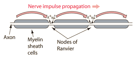
Transmission of a nerve impulse along an axonA nerve cell is like a receiver, transmitter and transmission line with the task of passing a signal along from its dendrites to the axon terminal bundle. The stimulus triggers an action potential in the cell membrane of the nerve cell, and that action potential provides the stimulus for a neighboring segment of the cell membrane. When the propagating action potential reaches the axon, it proceeds down that "transmission line" by successive excitation of segments of the axon membrane. Just the successive stimulation of action potentials would result in slow signal transmission down the axon. The propagation speed is considerably increased by the action of the myelin sheath. The myelin sheath around the axon prevents the gates on that part of the axon from opening and exchanging their ions with the outside environment. There are gaps between the myelin sheath cells known as the Nodes of Ranvier. At those uncovered areas of the axon membrane, the ion exchange necessary for the production of an action potential can take place. The action potential at one node is sufficient to excite a response at the next node, so the nerve signal can propagate faster by these discrete jumps than by the continuous propagation of depolarization/repolarization along the membrane. This enhanced signal transmission is called saltatory conduction (from the Latin saltare, to jump or hop). Tuzynski and Dixon offer some quantification of the sizes involved in these nerve cells. The axon is made up of connected segments of length about 2 mm and diameter typically 20 mm. This diameter compares to about 100 mm for the diameter of a human hair. Axon diameters may vary from 0.1 mm to 20 mm and may be up to a meter long. The much-studied squid has a giant axon of about a millimeter in diameter. The myelin sheaths are about 1mm in length. The action potential travels along the axon at speeds from 1 to 100 m/s. Contributing author: Ka Xiong Charand |
Index Bioelectricty Tuzynski & Dixon Sec 20.2 | ||
|
Go Back |