Heart Electrical Phenomena

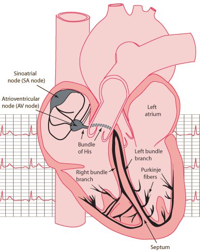
The rhythmic contractions of the heart which pump the life-giving blood occur in response to periodic electrical control pulse sequences. The natural pacemaker is a specialized bundle of nerve fibers called the sinoatrial node (SA node). Nerve cells are capable of producing electrical impulses called action potentials. The bundle of active cells in the SA node trigger a sequence of electrical events in the heart which controls the orderly pattern of muscle contractions that pumps the blood out of the heart.
The electrical potentials (voltages) that are generated in the body have their origin in membrane potentials where differences in the concentrations of positive and negative ions give a localized separation of charges. This charge separation is called polarization. Changes in voltage occur when some event triggers a depolarization of a membrane, and also upon the repolarization of the membrane. The depolarization and repolarization of the SA node and the other elements of the heart's electical system produce a strong pattern of voltage change which can be measured with electrodes on the skin. Voltage measurements on the skin of the chest are called an electrocardiogram or ECG.

The heart's electrical control system must properly synchronize the pumping functions illustrated above.
| Electrocardiogram |
Contributing author: Ka Xiong Charand.
Bioelectricty
| HyperPhysics***** Biology | R Nave |